What Can You Interpret From A Retinal Photograph?
While up to 90% of all vision loss is preventable, many eye diseases and conditions have few or no early symptoms, meaning that many people don’t realise that anything is amiss until their eyesight starts to change or an optometrist detects subtle changes through a retinal photograph.
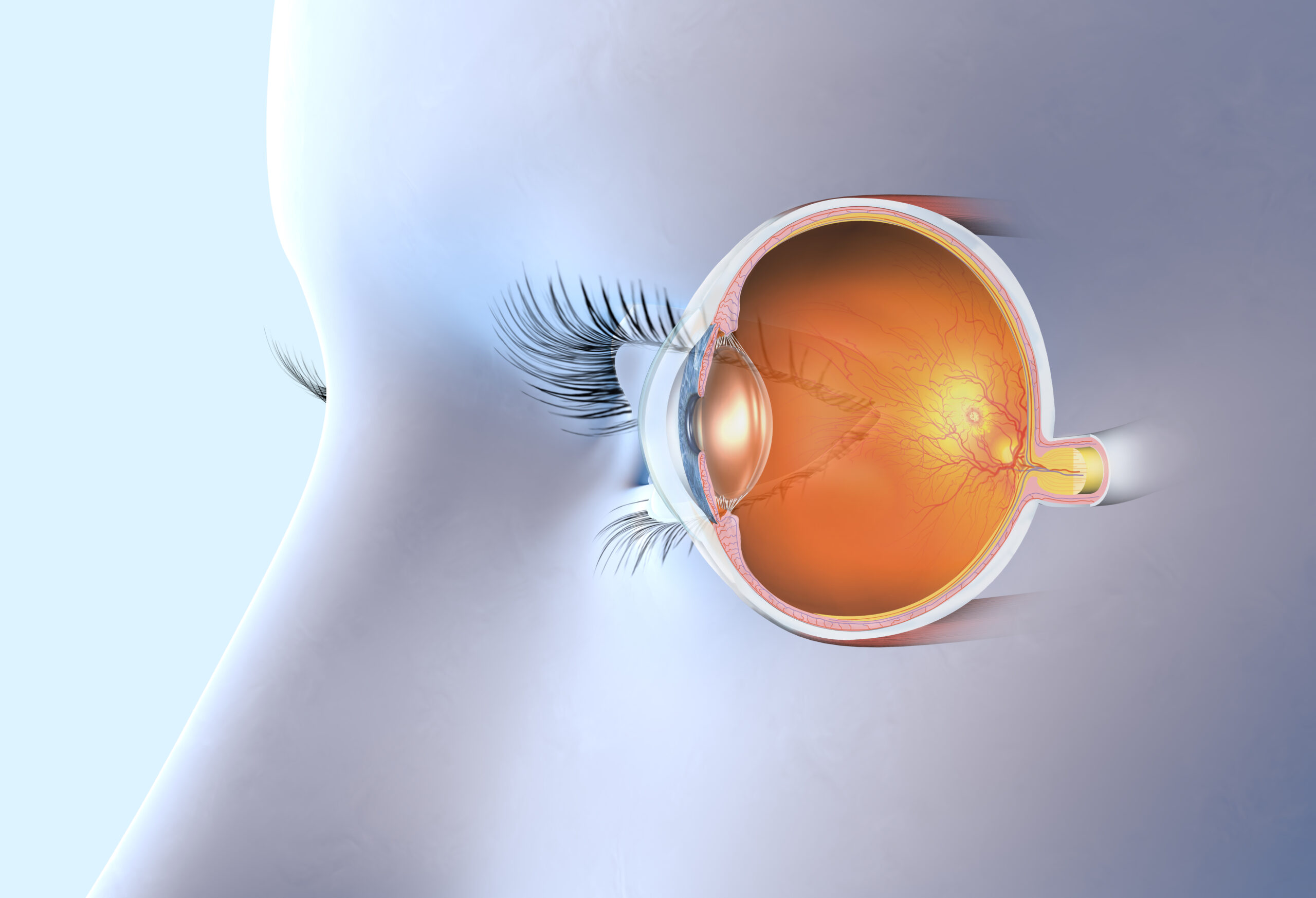
No other organ can be viewed in this way!
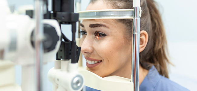
Early diagnosis and referral or treatment is therefore possible by using this technology, which is just one of the digital tools that optometrists at the Optical Company use. This high resolution camera allows one to observe the functioning of blood vessels, nerves, and tissues, without any invasive procedure.
The health of the eyes can in some cases, not only reveal potential eye conditions such as macular degeneration, glaucoma, haemorrhages and more, it can also be an indication of other systemic conditions. Examples of these are diabetes, high blood pressure , to name only two.
This screening process can help protect one of your most valuable assets, sight.
The retina is comprised of layers of extremely sensitive tissue at the back of the eye. In a perfectly healthy eye, nerves carry the messages received by the light reaching these membranes, to the brain.
How often is this necessary?
A full eye exam is recommended every 2 years in Australia if you are under 65, and annually if over 65. More regular checks will be recommended if any eye or systemic condition that affects eyes, including side effects of some medications, is present.
Digital photos that are saved can be compared in future years for monitoring and comparison.
Other digital occular screening devices include Optical Coherence Tomography (OCT), fundus photography and angiography.
Some examples of conditions that Optometrists can interpret from a retinal screening:
1. Diabetic Retinopathy
Diabetic retinopathy is a condition in which the blood vessels in the retinal become damaged by consistently high blood sugar levels, and it is a complication often seen in patients with poorly controlled diabetes.
In a retinal photograph, diabetic retinopathy will present with symptoms such as:
- Swollen veins in the retina
- Bleeding in the vitreous, the normally-clear gel that fills your eye
- New or abnormal blood vessels
- Possible scar tissue in advanced stages
- Possible retinal detachment in advanced stages, whereby the retina pulls away from its normal position at the back of the eye
2. Hypertensive Retinopathy
People with chronic hypertension, or high blood pressure, can develop hypertensive retinopathy, which can progress to loss of vision if left untreated.
The signs and symptoms of hypertensive retinopathy can include:
- Exudates or fatty deposits on the retina
- Bleeding in the retina
- White specks that resemble cotton wool, which indicate possible micro strokes
3. Retinal Tear and Detachment
On rare occasions, a retina may tear or even completely peel away or detach from the back of the eye. A detached retina is one of the more serious eye conditions and is considered a medical emergency, as it can lead to vision loss without urgent medical care.
Retinal tears and retinal detachments can indicate a range of conditions including age-related macular degeneration, eye injury and more, and occasionally retinal detachment can occur in the advanced stages of diabetic retinopathy.
4. Papilledema
Papilledema is the medical term for a swollen optic nerve due to increased pressure in the brain, which can indicate other underlying conditions such as:
- Very high blood pressure
- Eye injury or trauma
- Head injury or trauma
- Encephalitis, or inflammation of the brain
- Meningitis
- Stroke
- Tumours in the eye or brain
5. Optic Atrophy
Optic atrophy is the medical term for damage to the optic nerve, which is responsible for transporting images from the eye to the brain. Optic atrophy can be caused by medical conditions such as multiple sclerosis, stroke, cranial arteritis and tumours in the brain, and it can result in gradual vision loss over time.
Don’t delay treatment, get your eyes checked today. To book your comprehensive eye exam, with one of our experienced team members, contact one of your local clinics.
[1] https://www.visionaustralia.org/information/eye-health/eye-care